
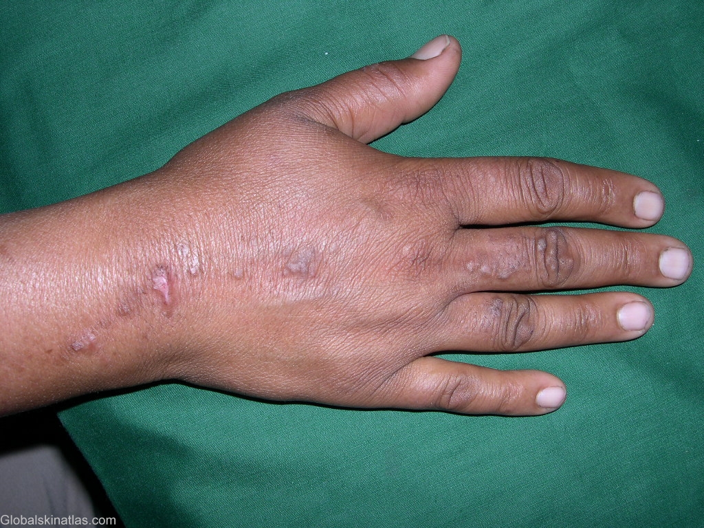
Diagnosis: Sporotrichosis
Description: Papulonodular lesions following the lymphatic pathway
Morphology: Abscess
Site: Hand,dorsum
Sex: F
Age: 50
Type: Clinical
Submitted By: Shahbaz Janjua
Differential DiagnosisHistory:
Sporotrichosis is caused by the dimorphic fungus Sporothrix schenckii, which is found throughout the world in decaying vegetation, sphagnum moss, and soil. The usual mode of infection is by cutaneous inoculation of the organism. Pulmonary and disseminated forms of infection, although uncommon, can occur when S. schenckii conidia are inhaled. Infections are most often sporadic and usually associated with trauma during the course of outdoor work. Infection can also be related to zoonotic spread from infected cats or scratches from digging animals, such as armadillos. Outbreaks have been well-described and often are traced back to activities that involved contaminated sphagnum moss, hay, or wood. Most cases of sporotrichosis are localized to the skin and subcutaneous tissues. Dissemination to osteoarticular structures and viscera is uncommon and appears to occur more often in patients who have a history of alcohol abuse or immunosuppression, especially AIDS. Spontaneous resolution of sporotrichosis is rare, and treatment is required for most patients. Although sporotrichosis localized to skin and subcutaneous tissues is readily treated, management of osteoarticular, other localized visceral, and disseminated forms of sporotrichosis is difficult.