
Diagnosis: Mycosis fungoides
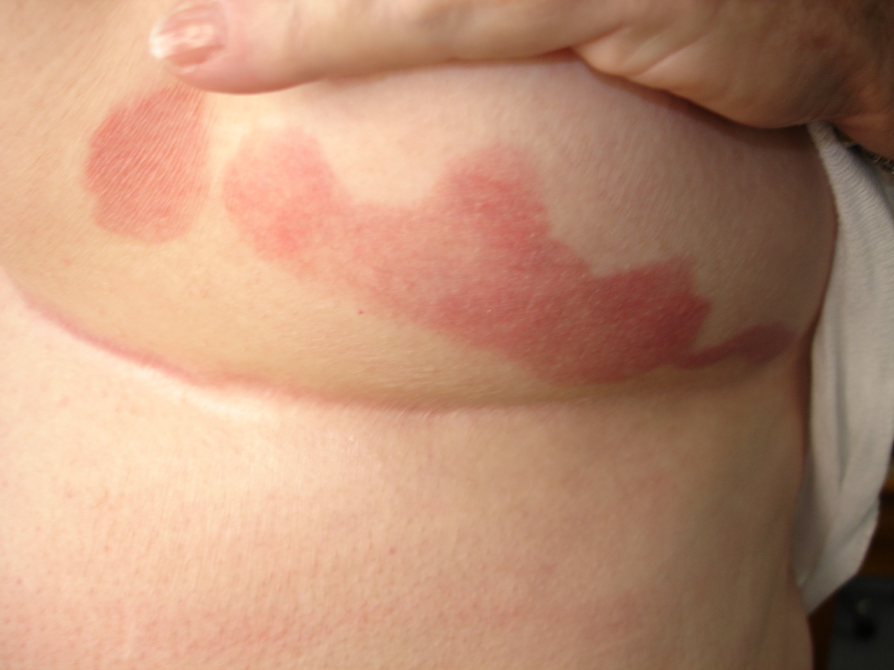
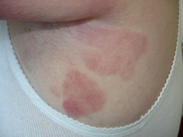
Description: Red non scaly patch persistent
Morphology: Red,nonscaly
Site: Breast
Sex: F
Age: 74
Type: Clinical
Submitted By: Ian McColl
Differential Diagnosis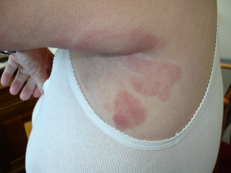
History:
|
The features of this lesion on the breast that should catch your eye are the straight line edge inferiorly, the overall sharpness of the edge and the curious tail of the lesion on the right side. Angulated edges like this are unusual in dermatology. One of the causes is mycosis fungoides or a T cell lymphoma of the skin. You might have considered psoriasis but it usually involves the breast flexure as does a sweating intertrigo. This lesion is so well defined it almost seems to have been painted on the skin. As the numbers of atypical lymphocytes in the skin increase this patch will become an itchy plaque and require radiotherapy. T cell lymphoma comes in many guises but this is a common presentation. The lesion is initially red and scaly and therefore comes into the differentials of the PMs PET (See Differential Diagnosis Blog ) The M of PMs PET is mycosis fungoides a T cell lymphoma of the skin but note that psoriasis, tinea and eczema are its differentials. An early patch of T cell lymphoma can be diagnosed as dermatitis by the pathologist because the characteristic intraepidermal changes have not be sampled properly by the biopsy. Hence you submit at least 3 punch biopsy samples from a suspected patch of T cell lymphoma and even then if the reported eczema does not respond to topical steroids you biopsy again! Progress Patches of T cell lymphoma can remain stable or grow very slowly for years without evolving into plaque or tumour stages but the condition is unpredictable. The earliest lesions are sometimes known as parapsoriasis both large patch and small patch parapsoriasis. They can be better diagnosed clinically than histologically. Patients can be seen with several lesional stages at once. Always biopsy the thickest lesion because there is more T cell infiltration there and you are most likely to pick up the Pautrier micro abscesses of atypical lymphocytes in the epidermis which are a hallmark of the condition. Investigations Multiple biopsies and a fresh specimen not in formalin for T cell receptor gene rearrangement studies using PCR or Southern blotting techniques.These should show a clone of T helper cells indicating the malignant nature of the lymphocytic infiltrate. Also do a full blood count and ask for Sezary cells in the blood plus the usual tests of liver and kidney function Treatment Try strong topical steroids on early lesions but refer early for UV therapy either PUVA or narrow band UVB. The latter though not penetrating into the skin as deeply as UVA light and oral psoralens, will do just as well for early patch and thin plaque lesions. It will not touch thick plaques or nodules. For those you need localised radiotherapy or systemic therapy including Interferon and oral retinoids, methotrexate and cyclophosphamide. |