Loading
Gloal Skin Path Atlas-logo
  • Recent Submissions
  • Search
  • Diagnosis
  • Quiz
  • Submit
  • Index
  • Blog
  • About GSA

<< Back to Diagnosis Page

Image Number #8549 (Sporotrichosis)

Diagnosis: Sporotrichosis

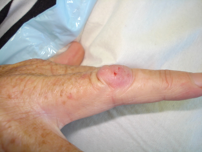
Description: Slow growing nodule on finger

Morphology: Nodule pink

Site: Finger

Sex: M

Age: 56

Type: Clinical

Submitted By: Ian McColl

View Full Size Image

Differential Diagnosis
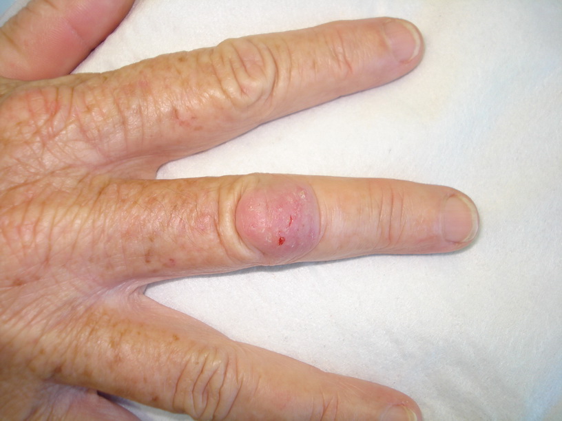
Click Here to View Details

History:

The patient has just finished a third course of antibiotics for a presumed infective swelling over the PIP joint of the middle finger with no real benefit. Do not bother prescribing a fourth. This swelling has been present now for 6 months and is more likely to be a deep fungal or atypical mycobacterial infection. Enquire about a fish tank at home for Mycobacterium marinum infection and whether the patient is a keen gardener for Sporotrichosis, a deep fungus found in old wood or mulch. Take a punch biopsy of the lesion for histology looking for granulomas and also send another specimen in an empty container for culture but specify for deep fungi and mycobacteria. The culture may take 6 weeks to grow.


Related Links:
Global Skinatlas on Sporotrichosis
Dermnet on Sporotrichosis
 

Case Comments     [Add Comment] [Subscribe]
This scenario is not uncommon. It is quite reasonable to view this lesion as infective or secondary to a foreign body reaction but you usually think of a bacterial infection as the likely infection and a splinter of wood as the foreign body. It is only when the second course of antibiotics fail that you sit back and wonder what is going on! We are used to fungal infections involving the epidermis giving a red lesion with a spreading peripheral scale but granulomas like this are due to fungal spores being introduced by injury under the skin and slowly "germinating" and growing. You then have a T helper cell response trying to wall off the infection and the lesion you see before you arises. (Submitted By: Dr Ian McColl)

    [Modify]

Common Deep Fungi The common deep fungi are sporotrichosis and chromoblastomycosis. Sporotrichosis is due to Sporothrix schenckii and is sometimes known as rose handler's disease because the organism is introduced from being pricked by a rose thorn. As well as forming a localised granuloma, if the person's immune response is impaired, the organism spreads up the lymphatics and may appear in the skin overlying the lymphatics as further granulomatous swellings. This lymphatic spread of sporotrichosis is so characteristic that it is known as sporotrichoid spread even when due to other organisms eg atypical mycobacteria from fish tank granuloma , but that is another story (Submitted By: Dr Ian McColl)

    [Modify]

Treatment Once you have cultured and identified the organism as sporotrichosis you can treat with a saturated solution of potassium iodide 3 drops tds increasing by a drop a day to 10 drops tds in orange juice to disguise the bitter taste. Cure may take three months! Other treatments include itraconazole 100-200 mgs daily for 3-6 months. This is available on the PBS for a proven deep fungal infection. Griseofulvin is useless! (Submitted By: Dr Ian McColl)

    [Modify]
 

Follow Up
View the relevant Module on this condition


Final Diagnosis

Deep fungal infection

DermNetNZ   eMedicine   PubMed   Dermatology Online   Archives   JAAD for "Sporotrichosis"

  • Recent Submissions
  • Search
  • Diagnosis
  • Quiz
  • Submit an Image
  • Index
  • About GSPA

Copyright 2022. Global Skin Atlas. ALL Rights Reserved.

Change Image