
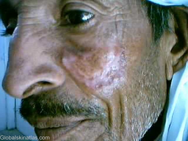
Diagnosis: Lupus vulgaris
Description: A large plaque with central scarring
Morphology: Plaque
Site: Face
Sex: M
Age: 60
Type: Clinical
Submitted By: Irfan Bari
Differential Diagnosis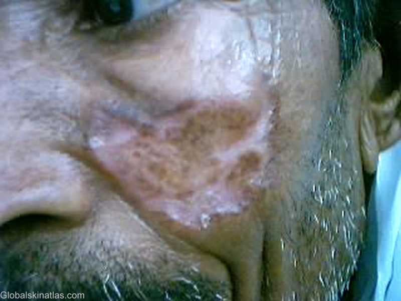
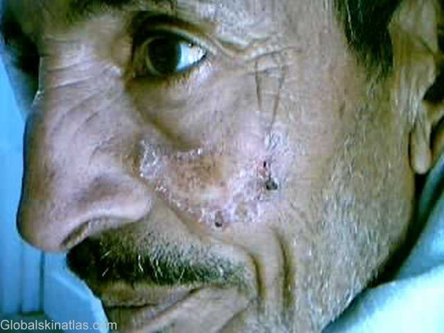

History: Lupus vulgaris is a chronic and progressive form of cutaneous TB that occurs in tuberculin-sensitive patients. In most series, it is the most common form of cutaneous TB.Lesions usually are solitary, and more than 90% involve the head and neck. Clinical variants are numerous and are seen as plaques, ulcers, nodules and vegetative forms. Histologically, the most prominent feature is a typical granulomatous tubercle with epithelioid cells, Langhans giant cells, and a mononuclear infiltrate. Caseation necrosis is minimal, and acid-fast bacilli are rare. Treatment regimens adequate for pulmonary TB also are effective for cutaneous disease. Cryotherapy is also effective. The role of surgery is limited for hypertrophic and verrucous lesions of lupus vulgaris .