
Diagnosis: Mycosis fungoides
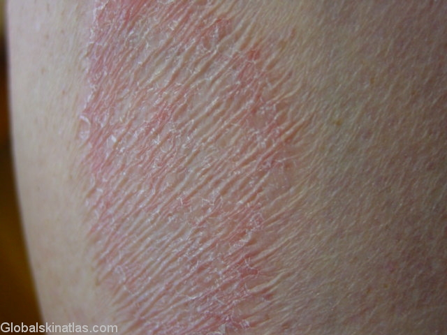
Description: Close up of plaque to show wrinkled surface
Morphology: Red,scaly
Site: Thigh
Sex: M
Age: 53
Type: Clinical
Submitted By: Ian McColl
Differential Diagnosis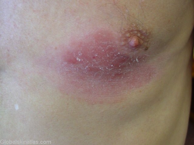
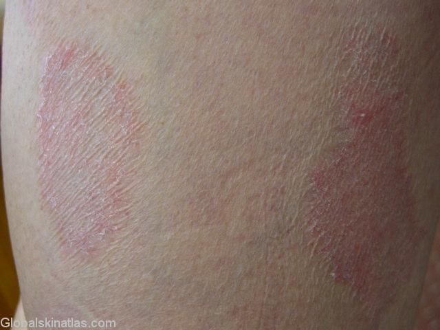
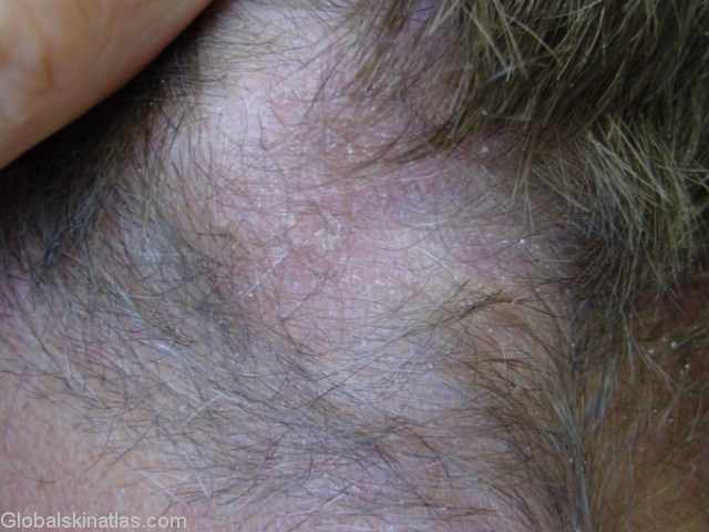
History: Mycosis fungoides does not really describe what we see above but this is the early morphological stage of T cell lymphoma of the skin.Skin associated T helper cells infiltrate the upper dermis and epidermis forming patches and then plaques with this characteristic wrinkling of the epidermis.Lesions at this stage will respond to both topical steroids and ultraviolet light therapy,both PUVA and narrow band UVB.The condition can be controlled for years with this therapy.Involvement of the scalp causes hair loss.If seen on its own it makes a clinical diagnosis of T cell lymphoma difficult!