

Diagnosis: Lupus vulgaris
Description: Red brown infiltrated plaque on the face
Morphology: Plaque
Site: Cheek
Sex: F
Age: 47
Type: Clinical
Submitted By: Ian McColl
Differential Diagnosis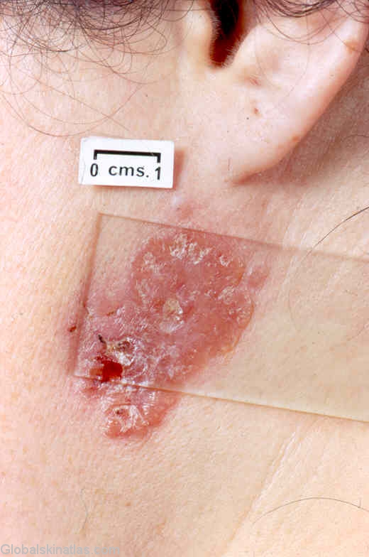
History: Lupus vulgaris typically occurs as a single plaque made up of red brown papules which show the characteristic apple jelly colour on diascopy.Most patients have moderately high immunity and will have a positive tuberculin test.Because of this immunity healing may occur in one area with new extensions at another.90% of lesions will occur on the head and neck.They can heal with considerable scarring.