
Diagnosis: Porokeratosis
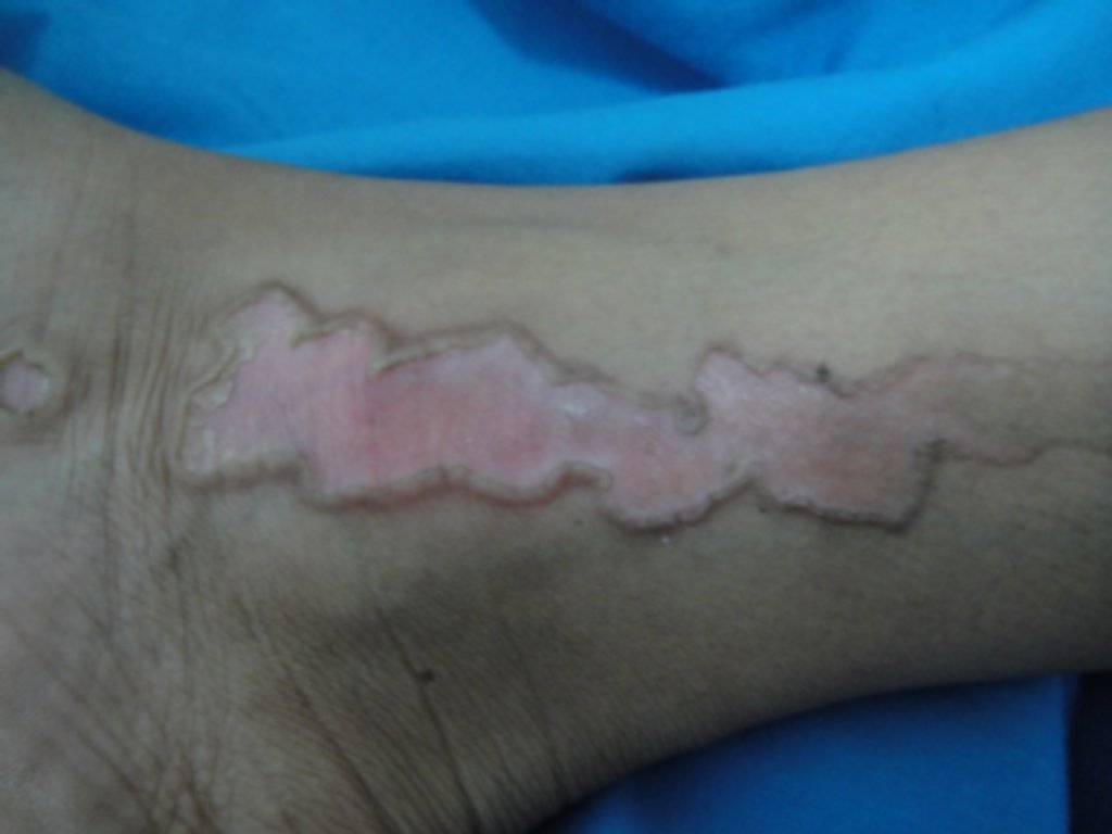
Description: Atrophic plaque with raised border.
Morphology: Atrophy
Site: Leg
Sex: F
Age: 24
Type: Clinical
Submitted By: Khalil Al-Hamdi
Differential Diagnosis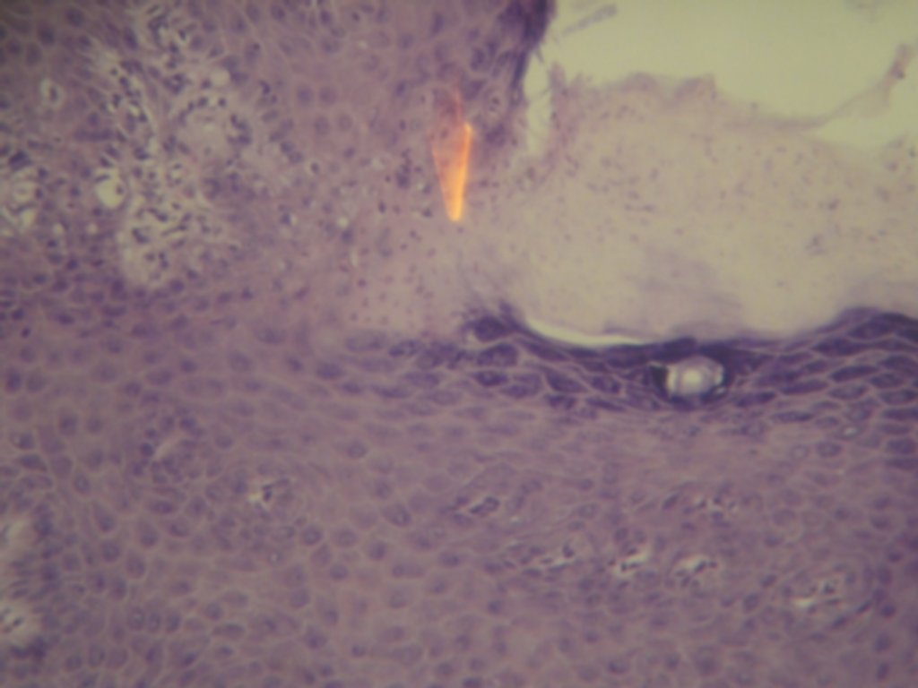
History:
A very distinctive and interesting case of unilateral linear porokeratosis presented with asymptomatic, linear, red, atrophic, scaly plaques with firm raised borders confined to one leg of one year duration. Skin biopsy taken from the border showed the characteristic and pathognomonic cornoid lamella i.e. a focal area of disruption of the granular layer with a column of parakeratotic cells in the stratum corneum (See the hisopathologic slide).