
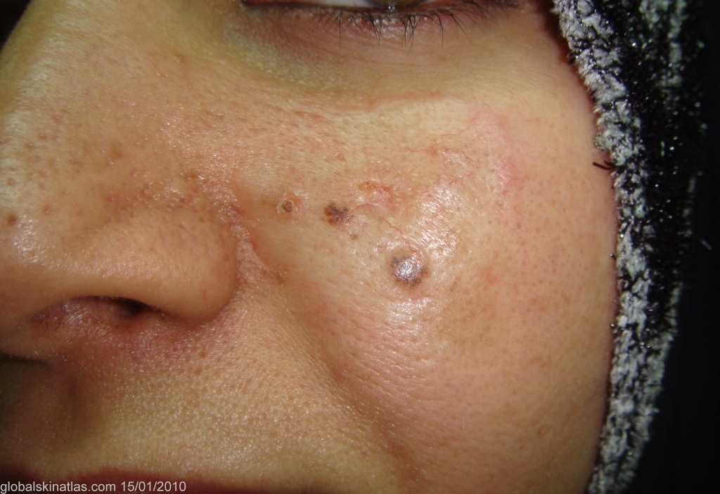
Diagnosis: Basal cell carcinoma
Description: Whitish sclerosed plaque.
Morphology: Sclerosis
Site: Cheek
Sex: F
Age: 37
Type: Clinical
Submitted By: Nameer Al-Sudany
Differential Diagnosis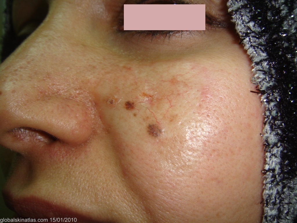
History: Morpheic or morpheaform BCC constitutes about 2% to 6% of all BCCs. It usually has a scar-like appearance hence its other name is cicatricial BCC. This middle-aged lady presented with a white sclerotic plaque on the left cheek with ill-defined borders of about 18 months duration. Surface telangectasia and some brownish black pigmentation were evident on examination. Incisional biopsy showed many strands of basal cells interspersed amid densely packed connective tissue.