
Diagnosis: Basal cell carcinoma
Description: This case illustrates a variant that has been left for years.
Morphology: Plaque
Site: Back
Sex: M
Age: 68
Type: Clinical
Submitted By: Ian McColl
Differential Diagnosis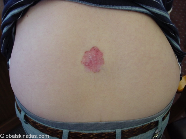
History:
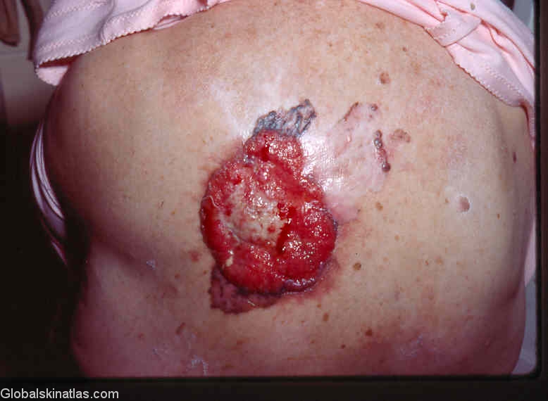
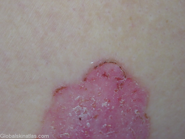
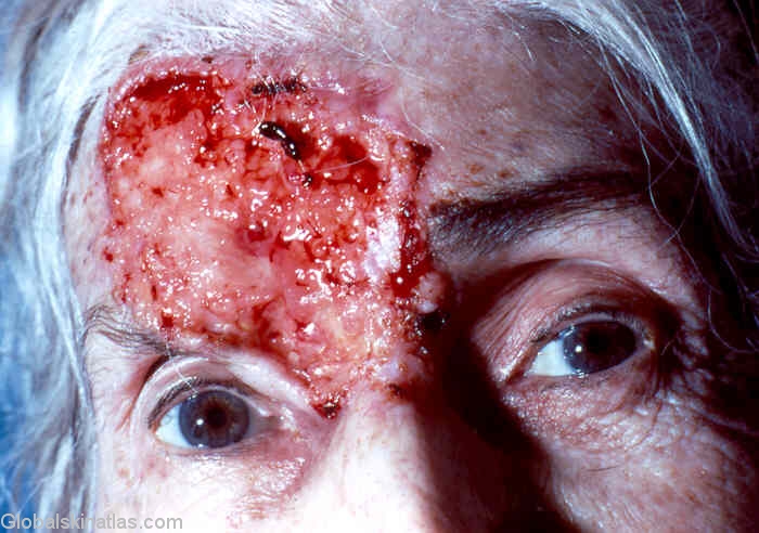
These four images show the spectrum of basal cell skin cancers.The superficial bcc grows as a flat scaly plaque for ten or more years to reach this size.The exophytic red fungating lesion on the back has not been adequately treated earlier but histology showed it was still a basal cell skin cancer.The facial lesion is similar.It has been left untreated for years.
BCCs usually invade tissues locally but do not metastasize elsewhere although these two neglected cases may spread to the local lymph glands.