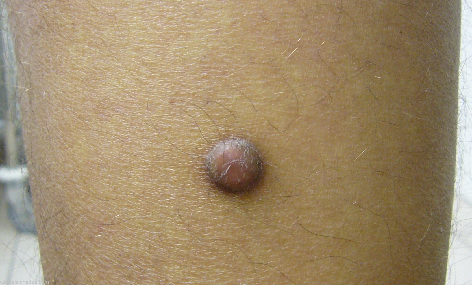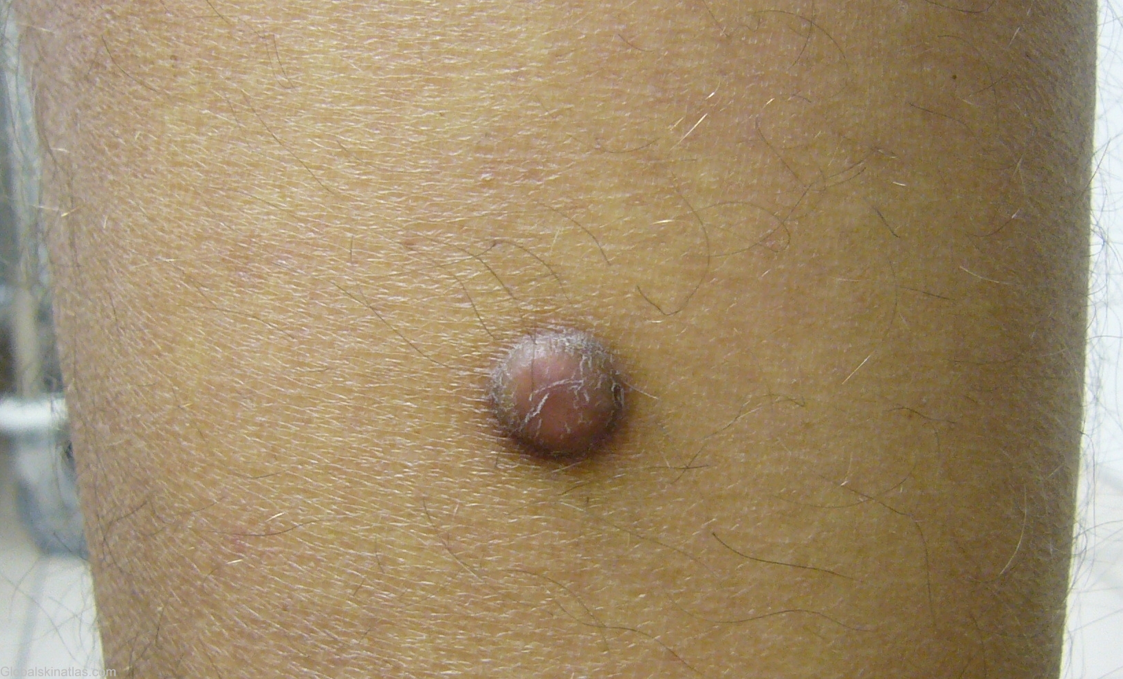

Diagnosis: Dermatofibroma
Description: 1 cm in size, dome shaped, red-brown in color, smooth surface and hyperpigmented ring surrounded.
Morphology: Nodule
Site: Calf
Sex: M
Age: 37
Type: Clinical
Submitted By: Mehravaran Mehrdad
Differential DiagnosisHistory:
Dermatofibroma Synonyms: fibrohistiocytoma, nodular subepidermal fibrosis, histiocytoma cutis and sclerosing hemangioma. Dermatofibroma is a common fibrohistiocytic tumor. It occur as papules or nodules on the extremities in approximately 80% of cases. The legs of women are a common location, possibly as a result of shaving or other minor trauma, and there are reports of cases involving the palms and soles. Although they have been reported to occur at any age, dermatofibromas most frequently affect individuals in early to middle adult life. There is no racial predilection. Although usually solitary, in about 20% of patients multiple lesions will be found, and there are reports of generalized dermatofibromas, especially in immunosuppressed patients. Treatment is not required, unless a lesion grows to several centimeters in diameter, when simple surgical excision may be indicated.
Case A 37-year-old male with chief complaint of a nodular tumor on the left calf. Physically the lesion was 1 cm in size, dome shaped, red-brown in color, smooth surface and with a hyperpigmented ring surrounding it.
