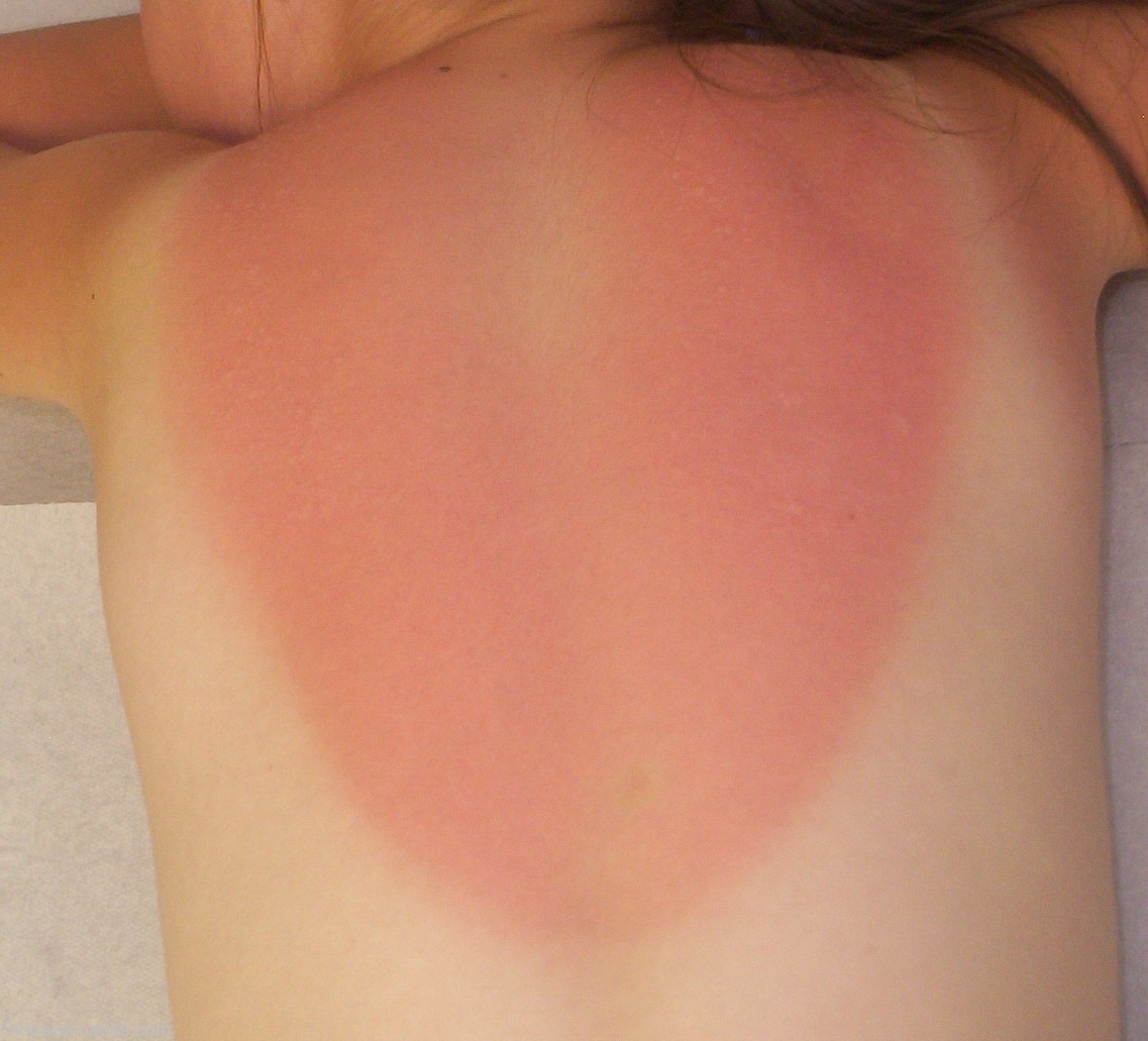

Diagnosis: Sunburn
Description: Diffuse, pruritic erythematous plaques on the sun-exposed areas.
Morphology: Photosensitive distribution
Site: Back
Sex: F
Age: 8
Type: Clinical
Submitted By: Mehravaran Mehrdad
Differential DiagnosisHistory: A first- degree burn of the skin results merely in an active congestion of the superficial blood vessels, causing an erythema that may be followed by epidermal desquamation (peeling). Ordinary sunburn is the most common example of the first-degree burn. The pain and increased surface heat may be severe, and it is not rare to have some constitutional reaction if the involved area is large. Light from the sun consists of three types of radiation, one of which is visible radiation, in the spectral range of 400 to 760 nm (violet through red). Beyond 760nm is infrared radiation, experianced as radiant heat. Below 400nm is the ultraviolet spectrum, divided in three bands. (UVA 320-390nm, UVB 290-320nm, UVC 230-290nm). The sun, whose rays contain 100 times more UVA than UVB during the mid-day hours, still burns us with just UVB. Sunburn is a normal human reaction to sunlight in excess of an erythema dose: i.e., the amount that will induce reddening (after a latent period of 4-8 hours) in an individual’s skin. The initial evidence is reddening, then soreness, and finally, is severe cases, blistering, which may become confluent. Discomfort may be severe; edema commonly occurs in the extremities and face; chills, fever, nausea, shock. In severe cases such symptoms may last for as long as a week. As the redness subsides and the blisters collapse, desquamation begins. A 8-year-old girl with diffuse, pruritic erythematous plaques with scattered small blisteres on the sun-exposed areas of the patient especially the upper back, which happened during swimming activity at midday. Meanwhile patient is under systemic antihistamine and local therapy.