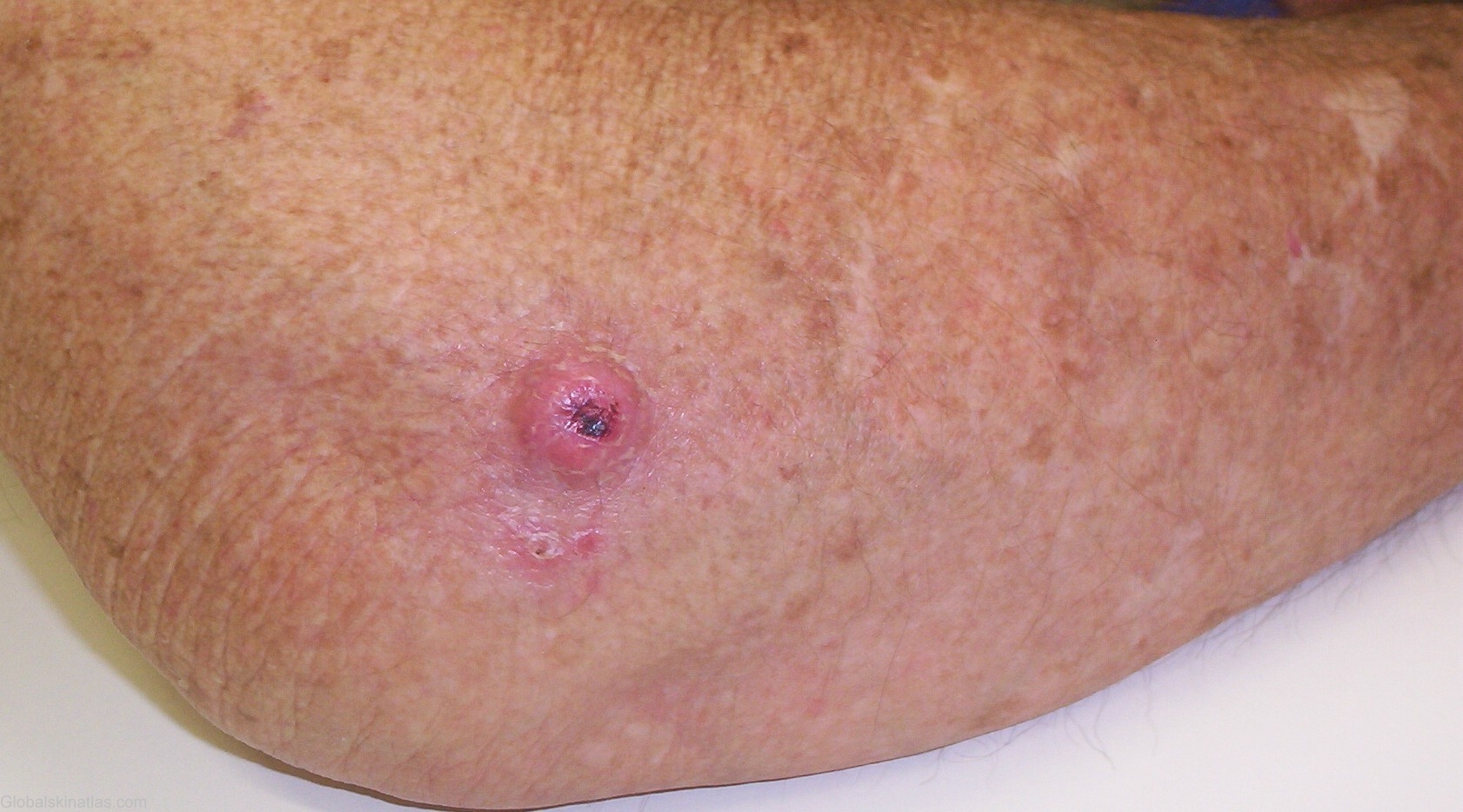

Diagnosis: Keratoacanthoma
Description: Single dome-shaped, skin-colored nodule in which there is a smooth crater filled with a central keratin plug.
Morphology: Nodule
Site: Elbow
Sex: M
Age: 76
Type: Clinical
Submitted By: Mehravaran Mehrdad
Differential DiagnosisHistory: Keratoacanthoma (KA) Synonyms: Self-healing primary squamous carcinoma, molluscum sebaceum, benign keratoacanthoma and idiopathic cutaneous pseudoepitheliomatus heperplasia. Definition: This term describes a suddenly (rapidly) appearing epidermal tumor with some of the characteristics of an Suamous Cell Carcinoma (SCC) but which resolves after a short period. Classically considered a benign epithelial neoplasm, KA shares many clinical and histologic features with SCC, and indeed, some consider it to be a form of SCC that usually, but not invarialbly, involutes. General Features: KAs typically arise on sun-damaged, hair bearing, light-colored skin in mid-to late life. Males are much more common affected that females. Lesions have been rarely noted to occur on mucous membranes and other non-hair-bearing skin such as the lips, intraorally, and subungually. While usually painless, KAs in certain locations, such subungual and intraoral, are painful. The most interesting feature of this disease is the rapid growth for some 2-6 weeks, which is followed by a stationary period for anther 2-6 weeks, and finally a spontaneous involution for another 2-6 weeks to leave a slightly depressed scar. The stationary period and involuting phase are variable , some lesions may take six months to a year to completely resolve. It has been estimated that some 5% of treated lesions recur, and 10-15% progress to SCC. A 76-year-old male with 3 weeks history of fast growing tumor on the right elbow. In toto excision and histopathology were done. Pathology proved the KA.