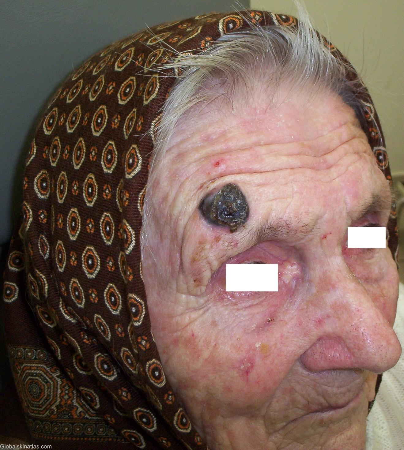

Diagnosis: Melanoma
Description:
Morphology: Path,tumour
Site: Forehead
Sex: F
Age: 90
Type: Clinical
Submitted By: Mehravaran Mehrdad
Differential DiagnosisHistory: Melanoma There are four recognized clinicohistologic classic types of melanoma: 1. Lenitigo maligna (melanoma in situ, noninvasive melanoma). 2. Superficially spreading melanoma (superficial spreading melanoma) 3. Nodular melanoma 4. Acral-lentiginous melanoma. Nodular Melanoma (NM) The typical lesion may be described as a pigmented papule or nodule of varing size, present for a few months. The lesions arise without a clinically apparent radial growth phase, but ususally large atypical melanocytes can be found in the epidermis for several rete redges beyond the region of vertical growth, at all margins of the excised lesion. NM constitues about 15% of all melanomas. It is twice as common in men as in women, and occurs primairly on sun-exposed areas of the head, neck and trunk. Although the tumors are at first 1-4 mm wide, they may grow much larger and become papillary, fungoid, or ulcerated. Bleeding is usually a late sign. The color is usually not uniform throughout the tumor but is likely to be scattered irregularly, being grayish brown, bluish-black, or black. A 90-year-old woman with chief complaint of broad, asymmetrical, unevenly pigmented patch/plaque with a nodule and a irregular border. ABCDs were very typical for NM. Patient was transfered to the regional melanoma center in Szeged for further management which includes in toto excision with saftey margin, staging, and further treatment.