
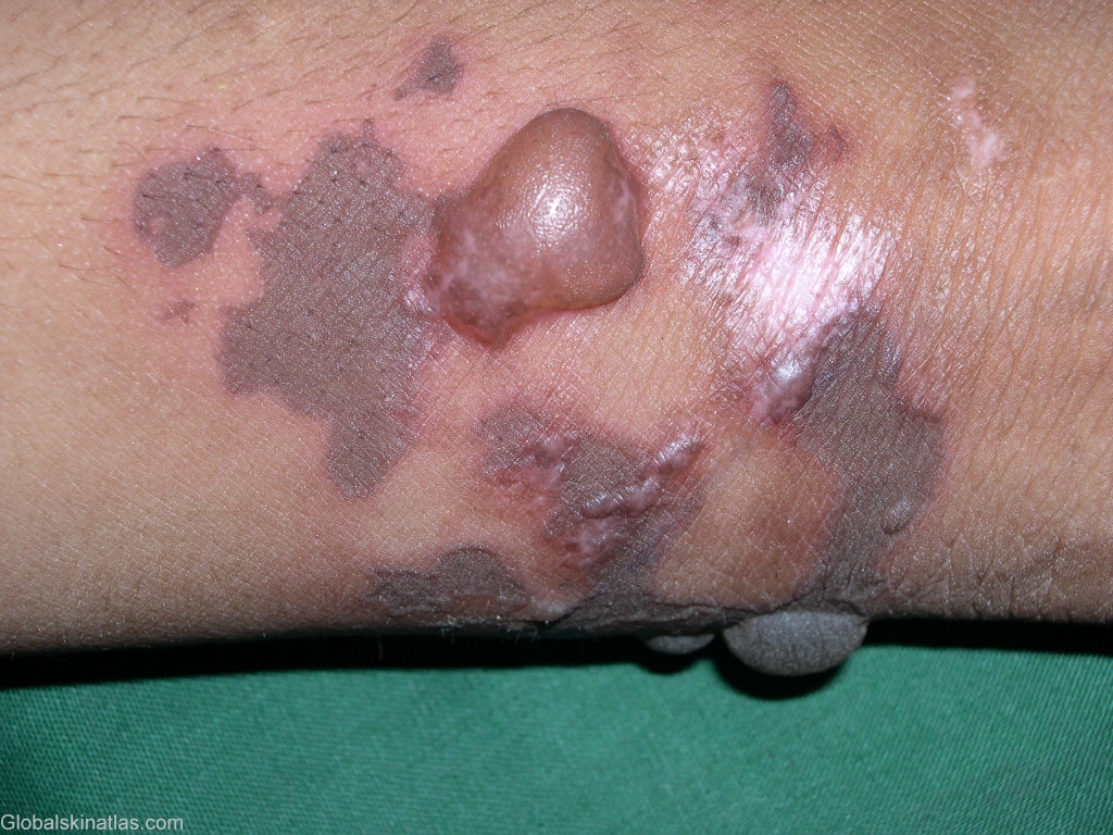
Diagnosis: Bullous fixed drug eruption
Description: Bullae and erythematous plaques
Morphology: Vesiculobullous
Site: Arm,forearm
Sex: F
Age: 34
Type: Clinical
Submitted By: Shahbaz Janjua
Differential Diagnosis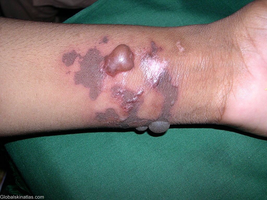
History: Fixed drug eruption (FDE) was first described by Brocq in 1894. The lesions of FDE usually start as an erythematous macule that subsequently evolves into a plaque. Vesicles and bullae develop at a later stage and are usually hemorrhagic. The lesions can occur on any part of the skin and mucous membranes. The sites of predilection are the limbs, sacral region, genitalia, palmar and plantar skin. The oral mucosa may be involved in association with skin lesions or alone.
The pathogenetic mechanism underlying FDE is still unknown . The most commonly accepted hypothesis is persistence of memory T cells in the affected skin. CD8+ cells phenotypically resembling effector memory T cells have been shown to be greatly enhanced in the lesions of FDE.