
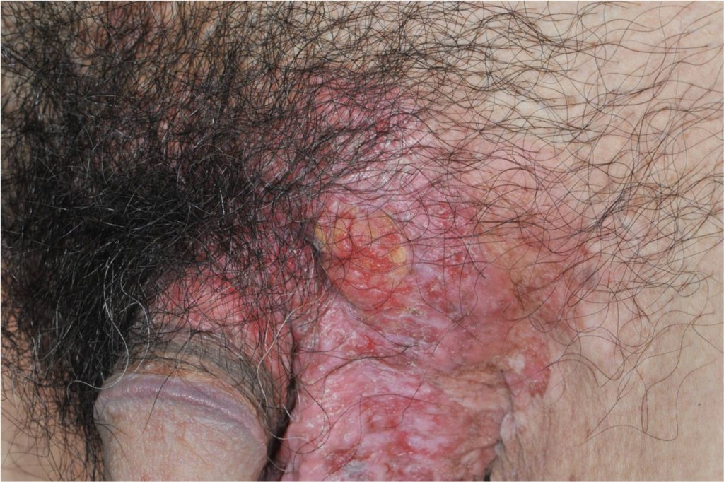
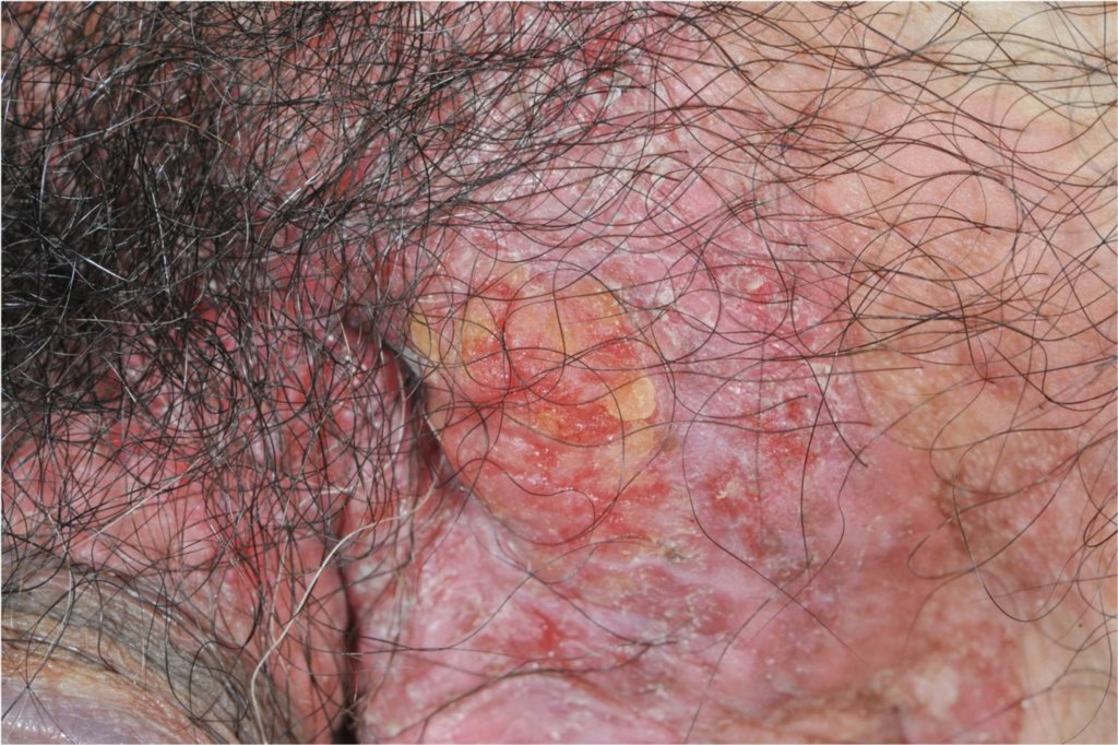
Diagnosis: Extramammary Paget's
Description: Eroded hyperplastic epidermis
Morphology: Erosions
Site: Scrotum
Sex: M
Age: 63
Type: Clinical
Submitted By: Ian McColl
Differential Diagnosis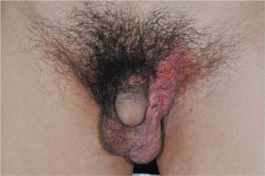
History:
Case of Dr
male 63 years old
Rash on left inguinal area for 5 months
Red, non-itchy, and scaly
Aggravated from sweating
Topical antifungal cream: not improved
Topical steroids cream: the lesion still persisted and spread more
No wt loss
Urological symptom : No urgency, no urinary dripping, no frequent voiding, no dysuria and no urethral discharge
GI symptom : No bowel habit change
No bone pain
Extramammary Paget’s disease (Favored Primary EMPD)
Secondary bacterial infection
KOH: Negative
Wood’s lamp: Negative
Gram stain : Mod. PMN, Numerous Gram + cocci
Skin Biopsy ? Incisional biopsy at Lt inguinal area
“Pagetoid” appearance
Extramammary Paget’s disease/ Paget’s disease
Pagetoid Melanoma
Pagetoid squamous cell carcinoma in situ
Mycosis fungoides
Cutaneous adnexal carcinomas (sebaceous carcinoma, porocarcinoma, and others),
Merkel cell carcinoma
Langerhans cell histiocytosis
other epidermotropic cutaneous metastases
The histological differential diagnoses you have given can all be correct! They need special immunoperoxoidase tests to determine the cell of origin. How did they treat him? (Submitted By: Dr Ian McColl)