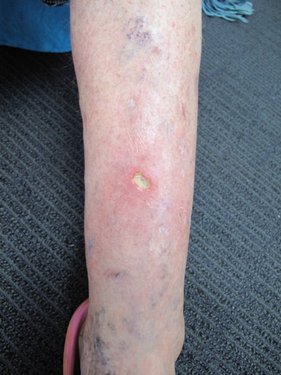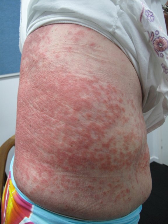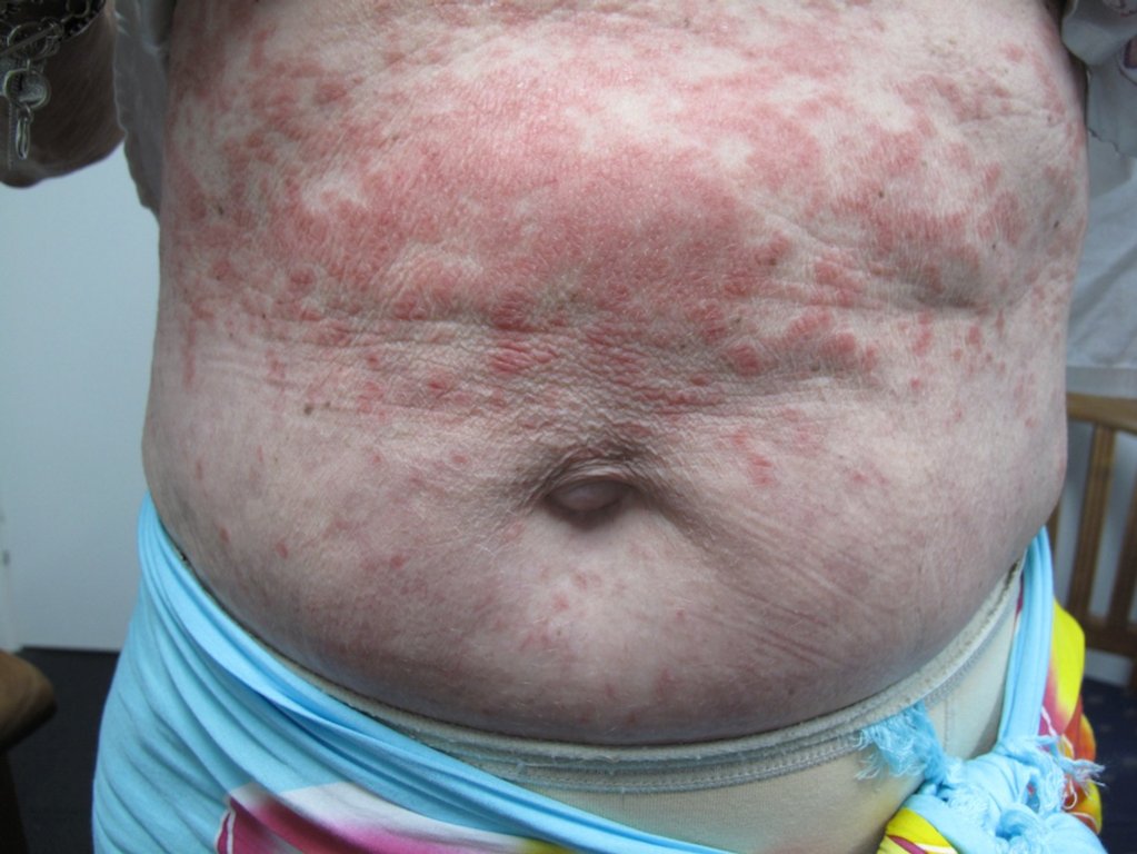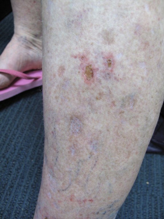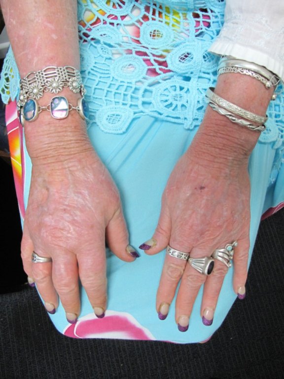
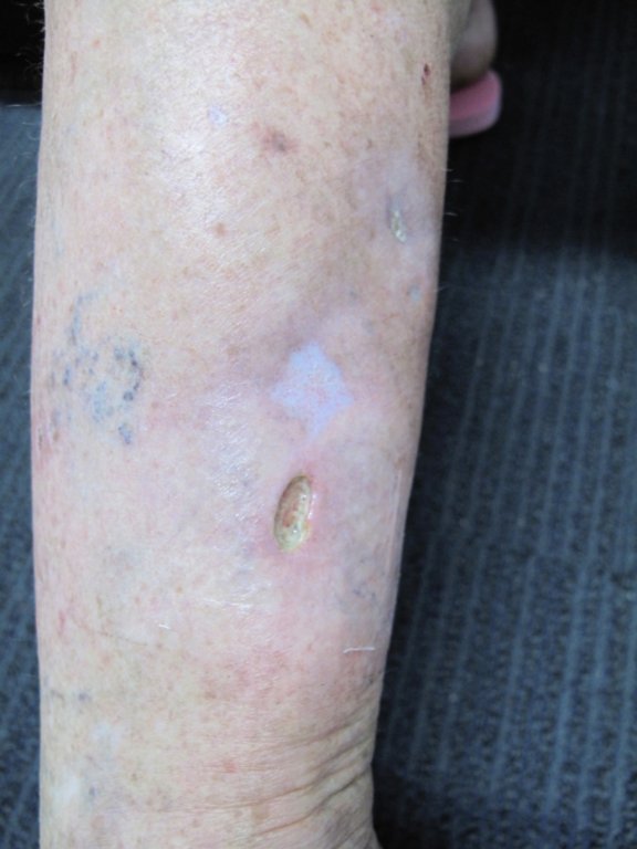
Diagnosis: Systemic lupus erythematosus
Description: Punched out ulcer
Morphology: Ulcer
Site: Shins
Sex: F
Age: 54
Type: Clinical
Submitted By: Ian McColl
Differential DiagnosisHistory:
Case of Dr Thomas Koeck This 54y old lady was diagnosed with SLE 7y ago.
When I saw her 4 months ago she was on 10mg pred.MTX 15mg and Hydroxychloroqine 400mg.
Also she took gabapentin for postherpetic neuralgia and a PPI,Adalat XL 30mg for Raynauds,Ramipril for kidney protection.and Naproxen prn for bad pain.She said she had a small MI 9y ago,but was not on the usual tablets.
Unfortunately she is not compliant with strict UV protection Despite the misery she is always cheerful!
I post this case for her shin leg ulcer. One on the posterior calf is healing slowly but the one over the lower ant shin is getting worse and now 13x20mm (the photo is older when it was smaller. Her ABI is 1.0.
Good periph pulses. She has some venous issues but cant tolerate compression as too painful. She had hives in the past and I think she has some urticarial vasculitis that looks like purpura.
THe rheumatologist thinks the ulcers are due to prednisolone (which she increases herself when her hands swell up or the skin rash is bad) SHe had a 50mg course scaling down to 5mg now but the ulcer got worse,not better. We tried Silversulfadiazine and alginate,not much impact though. Pseudomonas in the swab was rx w Cipro when she c/o severe pain and more redness around the ulcer but again no impact.
She smokes and doesnt put the legs up often. Actually hanging down is less painful,so I think the microcirculation is bad and compression is risky. I think ibuprofen flared her lupus.
ANA 2560 ,nuclear dots, ds-DNA neg. antiphospholipid and lupus coagulant neg. crea fine but C4 40 (range 130-780)
HEr lupus rash settled w diprosone only (and during the high dose pred of course)
How would you treat the ulcer? biopsy? admission?
Histo summary : ulceration anterior shin consistent with cutanous infarction. Lupus band test positive.Appearances are consistent with but not diagnositic of vasculitis.
DIF: small amount of IgG along the basal lamina of the epidermis.IgM is seen within the vessel wall showing fibrinoid necrosis and within the lumen of the larger artery showing intimal proliferation and luminal organisation.Complement and IgA are neg. (Submitted By: Dr Thomas Koeck)
She will need oral steroids and sometimes even cyclophosphamide. She needs to stop smoking! I would get a physician to see this one. She may infarct her kidneys or her brain! (Submitted By: Dr Ian McColl)
