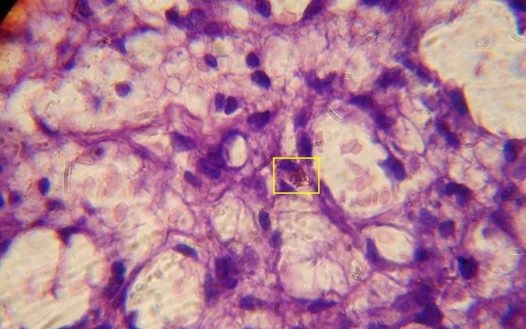

Diagnosis: Cutaneous Leishmaniasis
Description: Histopathology
Morphology: Nodule,purple
Site: Cheek
Sex: M
Age: 65
Type: Clinical
Submitted By: Nameer Al-Sudany
Differential DiagnosisHistory:
A 65-year-old man presented with two asymptomatic purplish-red nodules on his left cheek of two months duration. The lesions increased in size gradually. One lesion was excised totally and sent for histological examination. The lesion which was excised has recurred back after 3 weeks. On clinical background we can't definitely diagnose these lesions but as a differential diagnosis: lymphocytoma cutis, cutaneous leishmaniasis, pseudplymphoma, granuloma faciale, sarcoidosis, B-cell lymphoma, Jessner's Kanoff lymphocytic infiltrate, Bacillary angiomatosis and ALHE! The first reading of HP report by two histopathologist was histio-lymphocytic infiltrate with many abnormal lymphocytes and immunoblasts (B-cell lymphoma)!!! We didn't satisfy with histopathological report and it had been read by two dermatologists because so many histiocytes were a predominant histological finding! Surprisingly, on careful examination with high power lens we found so many parasitized histiocytes with Leishman-Donovan Bodies on H-E stain an evidence was enough to start our therapy. After 3-4 intralesional sessions of Gluconateme (Pentostam was not available at time of diagnosis) cure was obtained with no recurrence after two years follow-up! A case of Cutaneous leishmaniasis masqueraded as pseudolymphoma or even B-cell lymphoma! The messages obtained from this case:
(1) Query clinical cases should by proved by relevant investigation (biopsy) before taking any therapeutic decision!
(2) A clinico-pathological correlation is valid way to reach the final diagnosis in query cases
(3) Surgical excision is very rarely practiced in cutaneous leishmaniasis because the risk of scarring high recurrence rate!
