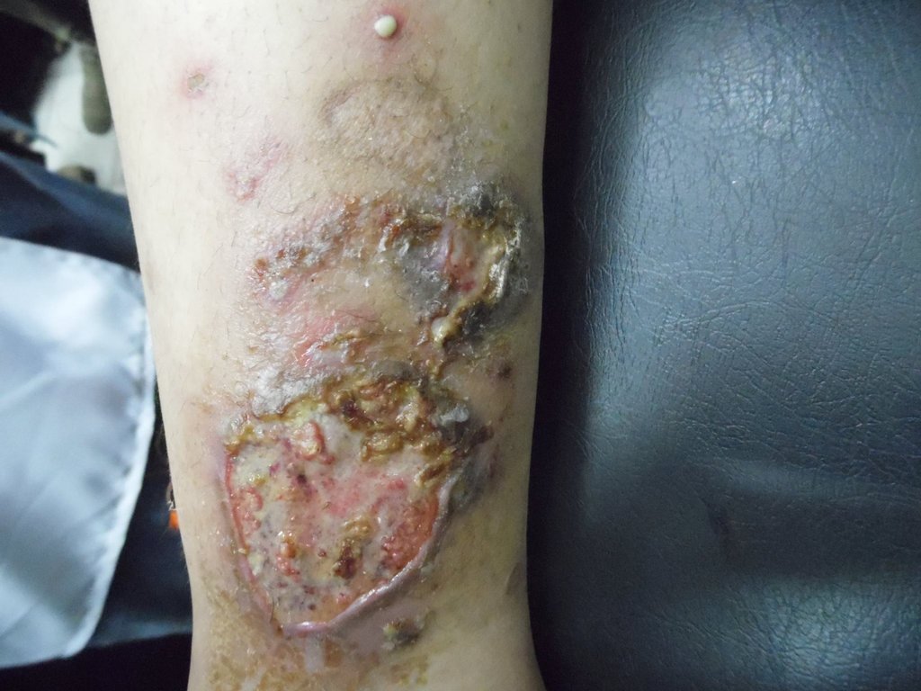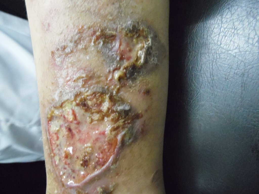

Diagnosis: Pyoderma gangrenosum
Description: Ulcers with undermined edges.
Morphology: Ulcer
Site: Leg
Sex: F
Age: 35
Type: Clinical
Submitted By: Nameer Al-Sudany
Differential DiagnosisHistory:
A 35-year-old woman presented with 6 months severely painful ulcer affecting her right leg. The patient reported current history of rheumatoid arthritis 5 years ago. Examination revealed; 2 ulcers 5*8 & 3*4 cm with evidence of atrophic scarring of the old lesion affecting the lower 1/3 of her right leg. The ulcer was severely painful with purulent base and dusky violaceous overhanging border. An intact 3 mm painful pustule seen in the vicinity of the lesion, the primary lesion. No response to repeated courses of antimicrobials prior to presentation. All laboratory findings were negative except for elevated ESR , CRP and iron deficiency anemia. Skin biopsy confirmed the diagnosis of pyoderma gangrenosum (sterile dense dermal neutrophilic infiltration (sea of neutrophiles)). The patient started on prednisolonoe 60 mg/d with dapsone 100 mg with proper dressing .
This case has been submitted by Dr. Ayman Abdelmaksoud, MD, Egypt
