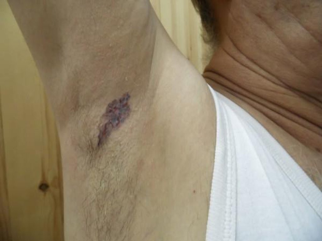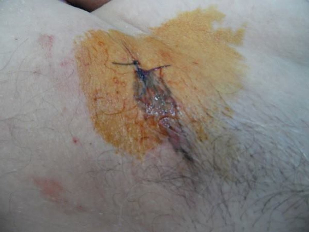

Diagnosis: Basal cell carcinoma
Description: Pigmented patch of long duration.
Morphology: Hyperpigmentation
Site: Axillae
Sex: M
Age: 68
Type: Clinical
Submitted By: Nameer Al-Sudany
Differential DiagnosisHistory:
Diabetic 68 years old military man presented with this pigmented patch that progressed slowly over the past 10 years on the right axillary fold. The lesion showed poor response to topical and systemic antibiotics. We put as a differential diagnosis:Melanoma...Superficial BCC..Pigmented and Bowen's disease.....The final diagnosis was Pigmented superficial BCC which usually characyerized by being more common in young age... involves maily the trunk and extremities and has fine thread like rolled border with eventually clearing hypopigmented center..The horizontal spread of the lesion is the role...Sub-clinical extension of the lesion is the cause of recurrence after surgical removal ,hence complete surgical excision with safety margin is favored by the treating oncologist.....What is new in this case; Older age (although elderly are more susceptible to Melanoma and non-melanoma skin cancer but this type is exception)...Superficial BCC shows variable degree of pigmentation, but the lesion here almost all pigmented that made confusion with the more serious melanoma....Axillae are not the common site for Superficial BCC.
This case is presented by Dr. Ayman Abdelmaksoud, MD, Egypt.

