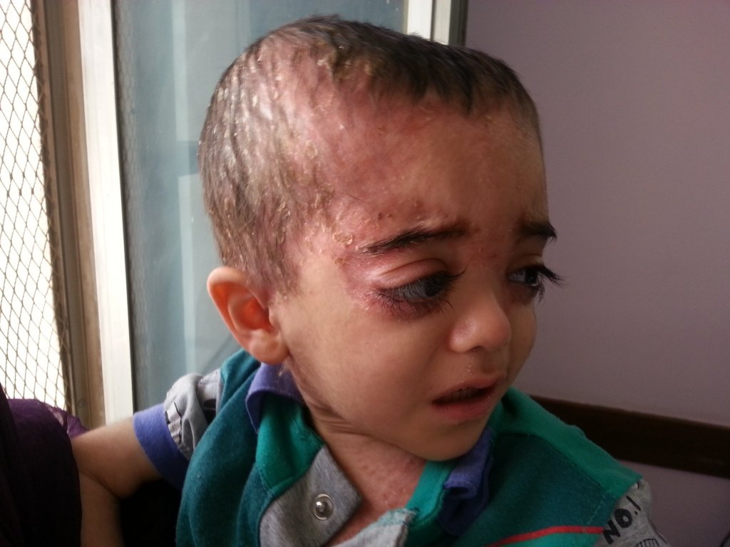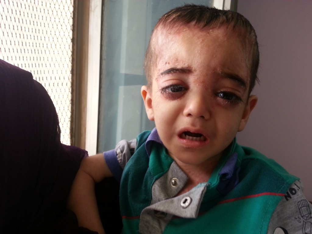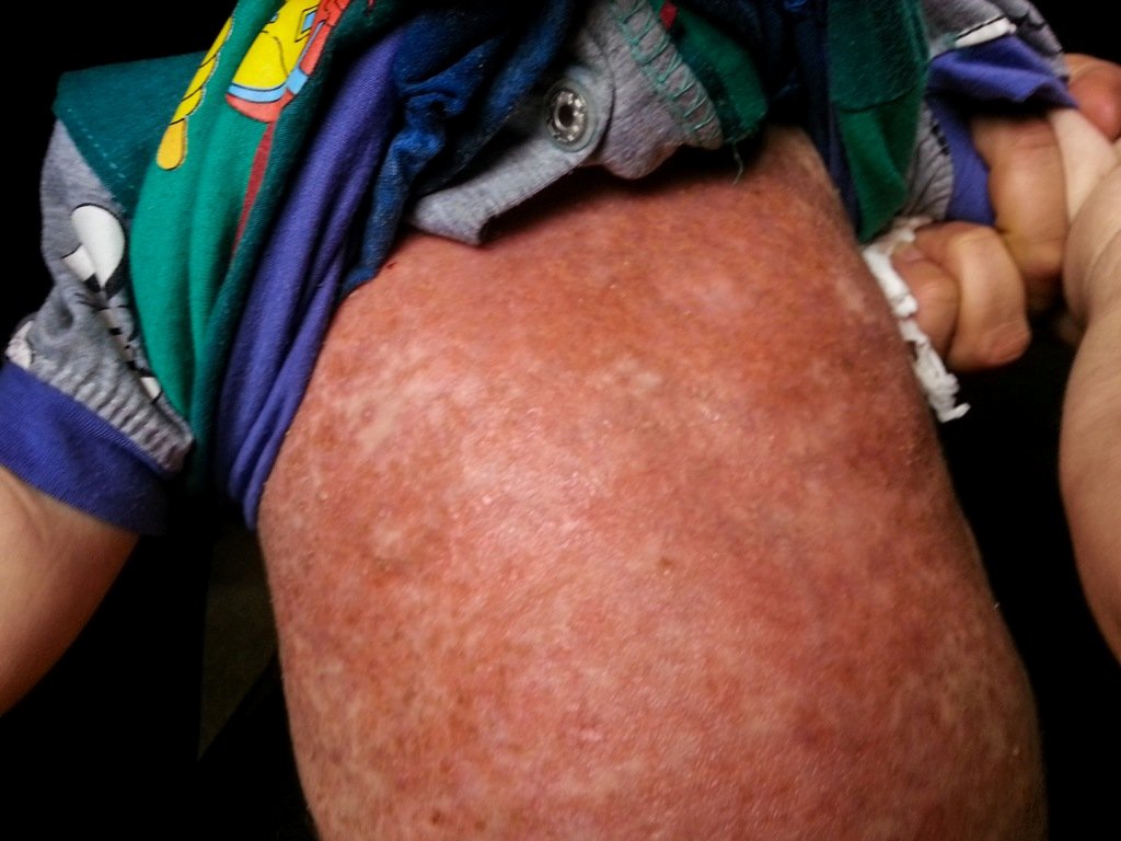

Diagnosis: Langerhans cell histiocytosis
Description: Scalp infections and asymmetrical protruding eyes.
Morphology: Red,scaly
Site: Face
Sex: M
Age: 4
Type: Clinical
Submitted By: Nameer Al-Sudany
Differential DiagnosisHistory:
A 4-year-old underweight male toddler presented with multiple scalp abscesses and red scaly rash involved the scalp, face and trunk of two years duration. O/E, hepatosplenomegally was found and many palpable painful lymph nodes specially in the cervical region were detected. The course was punctuated by relapses and partial remissions and some response to topical steroids and systemic and topical antibacterials was achieved on many occasions. Skull X-ray and Brain CT showed multiple osteolytic skull lesions. Many DDs were in mind at time of presentation:
1. Langerhans cell histiocytosis (LCH)
2. Hyper IgE syndrome (HIES, Job’s syndrome)
3. Leukemia and other lymphoproliferative disorders
4. Neuroblastoma with secondary metastasis
Clinically the presence of skull osteolytic lesions, hepatosplenomegally, seborrheic dermatitis-like rash made LCH is a priority in DDs.. Opinions of an ophthalmolosit and a neurologist were asked and neuroblastoma was excluded. Other investigations which had been arranged were CXR (Normal), CBC (All blood components were normal including WBCs differential count), IgG, IgA and IgE levels were normal. The final decision was made on skin and bone morrow biopsies, both of which proved the diagnosis of LCH. The child was admitted to a specialized pediatric oncology center and a course of chemotherapy (multi-organ involvement) was started. From the above work it clearly seems that management needs multidisciplinary team work (Dermatologist, pediatrician, radiologist, neurologist, ophthalmologist and oncologist) to deal with a multi-organ involvement case of LCH.

