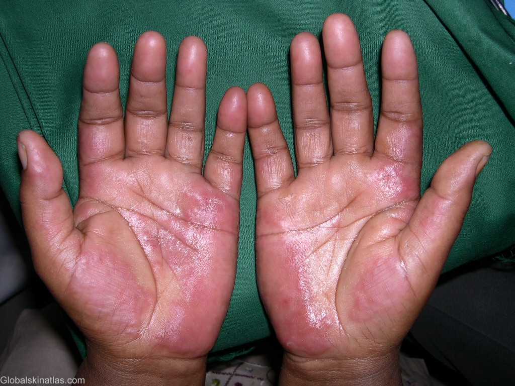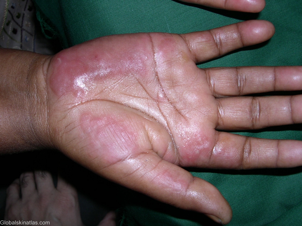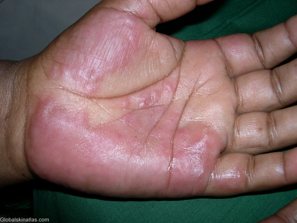

Diagnosis: Urticarial vasculitis
Description: Painful red non blanching plaques
Morphology: Red,nonscaly
Site: Hand,palm
Sex: M
Age: 14
Type: Clinical
Submitted By: Shahbaz Janjua
Differential DiagnosisHistory:
Urticarial vasculitis (UV) is an eruption of erythematous wheals that clinically resembles urticaria but is a form of leukocytoclastic vasculitis. UV plaques persist for 24 to 72 hours and may depict residual purpuric hues, scaling, and hyperpigmentation. Lesions are typically painful or burning rather than pruritic. UV is divided into two disease types termed normocomplementemic and hypocomplementemic. Both types can be associated with systemic symptoms (eg, angioedema, arthralgias, abdominal or chest pain, fever, pulmonary disease, renal disease, episcleritis, uveitis). The hypocomplementemic form more often is associated with systemic symptoms and has been linked to connective tissue disease (ie, systemic lupus erythematosus [SLE]). Direct immunofluorescence may reveal immunoglobulin and complement deposition in and around the blood vessel. UV should be suspected when individual painful hives persist for more than 24 hours or lead to pigmentation or show purpuric appearance. Poor response to antihistamines and features of systemic inflammation are also supportive. Urticarial vasculitis tends to run a chronic course. Antihistamines or nonsteroidal anti-inflammatory drugs (NSAIDs) may provide symptomatic relief in patients with only cutaneous involvement . If these agents do not work, colchicine, hydroxychloroquine, or dapsone may be prescribed. If all other treatment modalities have failed or if the patient has systemic involvement, systemic glucocorticoids may be advised. Steroid sparing agents like azathioprine may be added.
International Teledermatology Blog on this case.

