
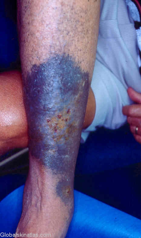
Diagnosis: Minocycline pigmentation
Description: Ulceration with surrounding blue black pigmentation due to treatment
Morphology: Hyperpigmentation
Site: Leg
Sex: M
Age: 62
Type: Clinical
Submitted By: Ian McColl
Differential Diagnosis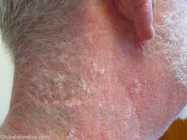
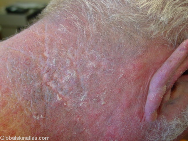
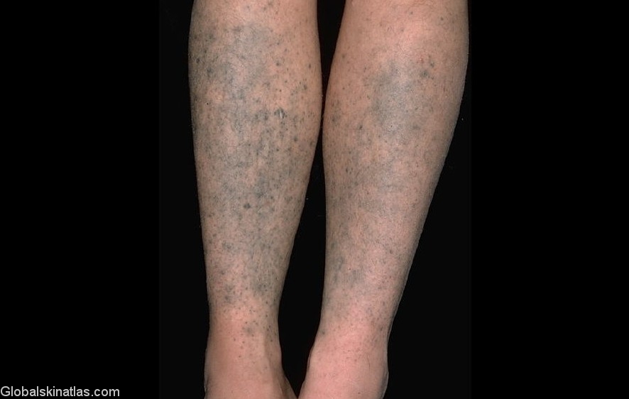
History: Minocycline pigmentation can present in three ways namely as pigmentation of acne scars,or as diffuse pigmentation of the lower limbs or as a diffuse generalised hyperpigmentation.The latter two presentations seem to be dose related but not the former.In addition to the skin Minocycline can be deposited in cartilage,conjunctivae,bone,sclerae and teeth.After stopping the drug it can take years for the pigmentation to fade.The leg ulceration being treated with Minocycline was caused by Pyoderma gangrenosum.