Loading
Gloal Skin Path Atlas-logo
  • Recent Submissions
  • Search
  • Diagnosis
  • Quiz
  • Submit
  • Index
  • Blog
  • About GSA

<< Back to Diagnosis Page

Image Number #816 (Reticular erythematous mucinosis)

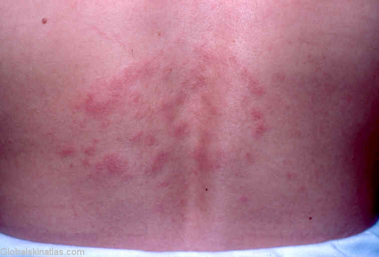
Diagnosis: Reticular erythematous mucinosis

Description: Close up of the infiltrates

Morphology: Annular

Site: Back

Sex: F

Age: 45

Type: Clinical

Submitted By: Ian McColl

View Full Size Image

Differential Diagnosis
Click Here to View Details

History:

Reticular erythematous mucinosis is a rare disorder typically seen in this area of the back,characterised by the deposition of mucin in the upper dermis.The condition is photoaggravated and is thought by some to be a variant of lupus erythematosus.

This lady partialy responded to topical steroids but cleared with hydroxychloroquine.

DermNetNZ   eMedicine   PubMed   Dermatology Online   Archives   JAAD for "Reticular erythematous mucinosis"

  • Recent Submissions
  • Search
  • Diagnosis
  • Quiz
  • Submit an Image
  • Index
  • About GSPA

Copyright 2022. Global Skin Atlas. ALL Rights Reserved.

Change Image