
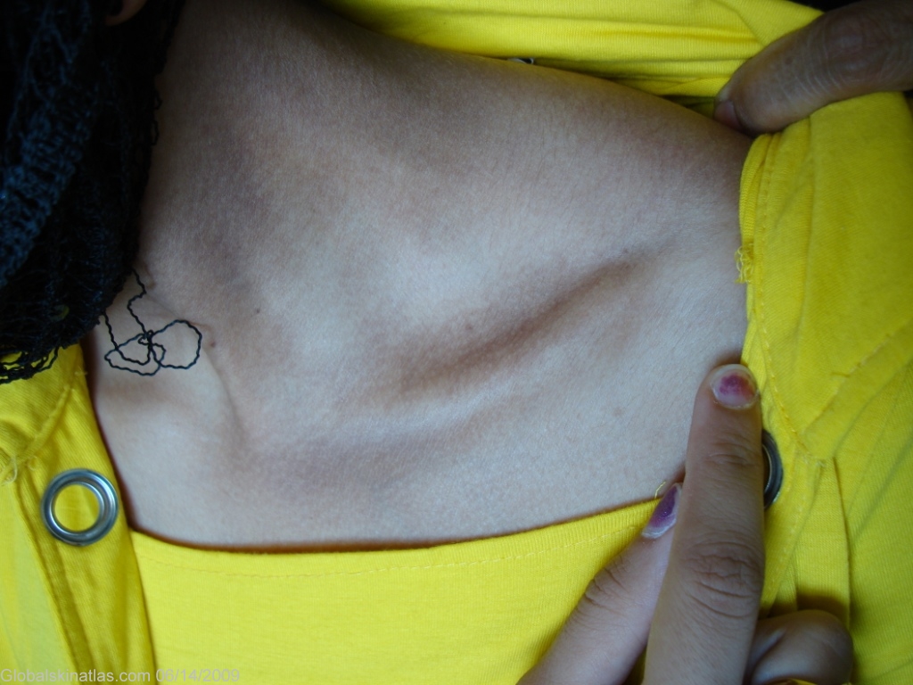
Diagnosis: Macular amyloidosis
Description: Rippled, hyperpigmented patches on the side of the neck.
Morphology: Hyperpigmentation
Site: Neck side
Sex: F
Age: 22
Type: Clinical
Submitted By: Nameer Al-Sudany
Differential Diagnosis
History:
Macular amyloidosis may be confined to the interscapular area but more commonly the lesions are more extensively distributed over the back, neck, chest and the extensor aspects of extremities. Macular amyloidosis has been reported to follow prolonged chronic friction, such as the use of a nylon brush during bathing. The skin lesions are hyperpigmented, although hypopigmented areas may also occur giving a 'poikilodermatous' appearance. A reticulate or 'rippled' pattern of pigmentation is a characteristic diagnostic feature in many cases of macular amyloidosis. The lesions tend to be associated with mild to moderate pruritus, but pruritus may be absent in some cases. The condition usually presents in early adult life and persists for many years; both sexes are equally affected.